Echocardiography FAQs
-
- Our team will provide any specific instructions at the time of booking, and these should be followed carefully.
- You will be asked about your cardiac history, such as valve replacement types and previous echocardiogram results. If you are able to, please bring these results and record with you.
- You may be asked to change into a gown during the procedure, so we recommend wearing separate top and bottoms during your appointment as this makes changing for the examination easier.
- Please do not apply any body lotion or sunscreen to your chest before the appointment, as we need to apply ECG patches.
- If you are pregnant, please let the technician know.
-
At the start of your appointment, your sonographer will explain exactly what will happen during your procedure. It may vary slightly from what we describe below, so don’t worry if this is the case; you’ll always be in very good hands.
- Before the scan, you will be asked to remove all clothing above the waist (including undergarments). Female patients will be provided with a gown to wear during the examination.
- Your blood pressure will be taken, and you will be asked to confirm your weight and height.
- You will be asked to lie on a bed, and small ECG patches will be applied to your chest; these are sticky “dots” with leads attached. ECG patches allow us to record your heart rhythm.
- The scan is performed lying on your left side, with your left arm above your head.
- The sonographer will apply a warm gel to your chest, which allows the transducer to move smoothly over the surface.
- The sonographer will then press the transducer against your skin and move it over your chest area.
- Throughout the examination, the sonographer will ask you to change positions and breathe in specific ways. This is so they can capture clear and accurate images of your heart from different angles. Please follow these instructions as best you can.
-
An echocardiogram can take from 20 to 45 minutes to complete; occasionally, however, the scan can take longer.
-
The results of your cardiac ultrasound will be sent directly to your referring doctor. Please ensure you book a follow-up appointment to discuss your results with them.
Queensland X-Ray provides patients with all of their images via our Patient Portal. Shortly after your first visit to Queensland X-Ray, you will receive a text message about accessing your results through our Patient Portal. This secure portal allows you to view and download your medical images and results at any time. It is compatible with most internet browsers and mobile devices, ensuring accessibility from anywhere in Australia.
-
An echocardiogram is a non-invasive procedure that is not painful, although you may experience some discomfort. During the exam, the sonographer will use a transducer to capture images of your heart, which involves applying pressure around the ribs and chest area. This area can be sensitive.
-
Yes, an echocardiogram is performed using ultrasound, which is a very safe imaging technique, with no known negative effects.
-
Your echocardiogram will be performed by a specially trained sonographer, known as a cardiac sonographer or echo sonographer.
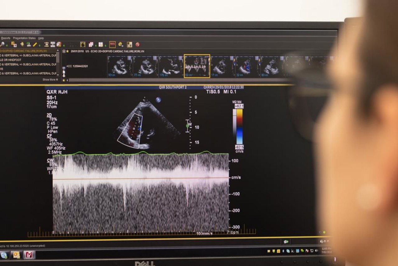
The images captured by the sonographer are viewed by a radiologist or cardiologist, who will provide a report of findings.
-
An echocardiogram or cardiac ultrasound provides important information needed to diagnose and manage heart conditions.
This scan can assess the size of the heart chambers, wall thickness, heart muscle function, progression of heart disease, valve function, masses in the heart, fluid around the heart, holes or defects, and abnormalities of blood flow within the heart. It can help diagnose atherosclerosis, cardiomyopathy, congenital heart disease, and cardiac tumours.
How much will it cost?
Fees for radiology procedures will vary depending on a variety of factors. We will advise you about the cost of your service at the time of booking but if you do have any questions, contact us and one of our team will be happy to help with your query. You can read more about our billing information here.
How do I access my images?
At Queensland X-Ray, we provide our patients with their images and results online. To access your images and results, you’ll need to register for an account when you visit one of our practices. If you’ve already registered, you can access the Patient Portal here.