PET imaging can be a useful tool for identifying specific regions of hypo metabolism in the brain which can help in the diagnosis of certain neurological conditions. PET imaging involves injecting a small amount of radioactive tracer into the body, which is taken up by the cells in the brain. The tracer emits positrons, which can be detected by the PET scanner and used to create images of the brain. By selecting a tracer that is taken up specifically by cells in the regions of interest, PET imaging can be used to identify areas of hypo metabolism or decreased activity in that region. This can be particularly useful in the diagnosis of conditions such as Alzheimer’s disease, where there is often hypo metabolism in the temporal and parietal lobes of the brain. PET imaging can also be used to monitor the progression of certain neurological conditions and to evaluate the effectiveness of treatments.
FDG PET Brain Scan FAQs
-
Our preparation includes a reproducible low carbohydrate diet that removes variables, meaning that your FDG PET scans will be more accurately compared from baseline scans through to post-treatment scans.
When making your appointment, a member of our clerical team will provide you with preparation instructions for your scan. This will include suggested foods to eat in the 24 hours leading up to your scan. Additionally, you will be required to fast from food for a minimum of six hours prior to your appointment. You will receive an appointment confirmation call on the day prior to your appointment where you can ask any questions you may have.
-
Arrival (15-30 minutes)
When you arrive for your appointment, our reception staff will ask that you complete a questionnaire and sign a consent form. In line with your appointment time a registered nurse will welcome you through for the procedure. Please be mindful that some patients take longer than others to get settled and your appointment time may vary.Tracer uptake (30 minutes)
A nurse will come and settle you into a private uptake room. They will explain the procedure, take your medical history and place an IV cannula into your arm. An IV line will be connected through your cannula, through which the tracer will be injected. At this point you will be left to sit in the dimmed uptake room with your eyes open whilst the tracer is infused. After approximately 30 minutes, staff will take you through to the PET/CT scanner.
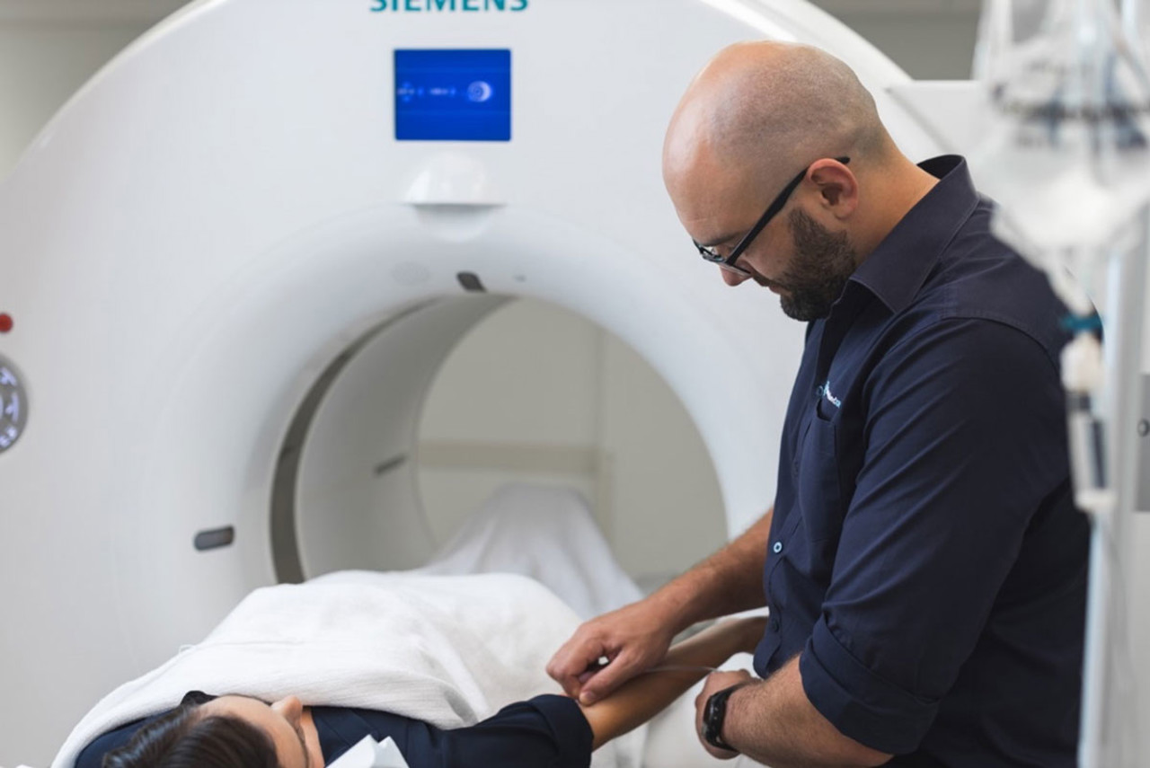
Any metal that you may have on your face (i.e jewellery, dentures) will need to be removed prior the scan.PET/CT imaging (15-20 minutes)
The PET/CT scanner looks like a doughnut (known technically as a gantry), with a bed passing through the middle. You will be asked to lie on your back, with your head placed in a head holder. Foam blocks and velcro straps are placed around your head to secure it from any movement.The PET part of the scan detects the radiation from the tracer which has now been absorbed into the brain. This illustrates physiological uptake.
The CT part of the scan images the structural components and anatomy of the brain.
The PET and CT images are fused together to create an image that highlights patterns of metabolism.
Recovery time (15-30 minutes)
Following your scan you will be offered something to eat and drink, be encouraged to stay well hydrated and to use the bathroom if required. We will monitor you for 15 minutes following the CT contrast injection and then remove your IV line. During this time, the staff will process your images. -
An FDG PET brain scan may take up to 3 hours to complete. Please refer to the above item 'what will happen during my appointment' for more information.
-
Queensland X-Ray has the latest nuclear medicine equipment, with PET/CT available in most regions.
A nuclear medicine technologist will perform your scan with support from a registered nurse, and under the supervision of a nuclear medicine specialist.
The images the technologist acquires are reviewed by a specialist who reports the findings. This report will be sent to your referring doctor who will explain the results to you. Please note that technologist(s) performing the scan do not discuss scan results.
-
Fees for PET/CT scans will vary depending on the type of pathology under investigation. We will advise you ahead of time about the cost of our service. However, if you have any questions, please feel free to contact us.
How much will it cost?
Fees for radiology procedures will vary depending on a variety of factors. We will advise you about the cost of your service at the time of booking but if you do have any questions, contact us and one of our team will be happy to help with your query. You can read more about our billing information here.
How do I access my images?
At Queensland X-Ray, we provide our patients with their images and results online. To access your images and results, you’ll need to register for an account when you visit one of our practices. If you’ve already registered, you can access the Patient Portal here.