Vascular Ultrasound
Vascular ultrasound imaging is a non-invasive ultrasound examination used to assess blood flow through the veins and arteries in different parts of the body. It uses high-frequency sound waves to evaluate how blood moves through blood vessels.
Vascular ultrasound imaging is commonly used to help assess circulation, identify changes in blood flow, and check for conditions affecting the body’s circulatory system.
Vascular Ultrasound FAQs
-
Your doctor may refer you for a vascular ultrasound to assess blood flow in your veins or arteries or to investigate symptoms that may be related to circulation. This examination is commonly requested when there is concern about reduced, altered, or absent blood flow.
Vascular ultrasound is often used to help assess conditions such as suspected blood clots, including deep venous thrombosis, identify blockage or narrowing of arteries, varicose veins, or changes in blood flow that may increase the risk of stroke or other vascular conditions.
-
A vascular ultrasound can help assess a range of conditions affecting the arteries and veins by evaluating blood flow and vessel structure. This form of vascular imaging may be used to help detect blood clots, areas of reduced or absent blood flow, narrowing or blockages in arteries, and abnormal connections between blood vessels, such as arteriovenous malformations.
Vascular ultrasound may also be used to assess varicose veins, dialysis fistulas, congenital vascular malformations, and other medical conditions affecting the body’s circulatory system. Findings from this examination help support clinical decision-making by your referring doctor.
-
- Our team will provide any specific instructions at the time of booking and these should be followed carefully.
- We recommend wearing a separate top and bottoms during your appointment as this makes changing for the examination easier.
-
You will be asked to lie down on a bed or stand and the part of your body we’re imaging will need to be visible throughout the examination.
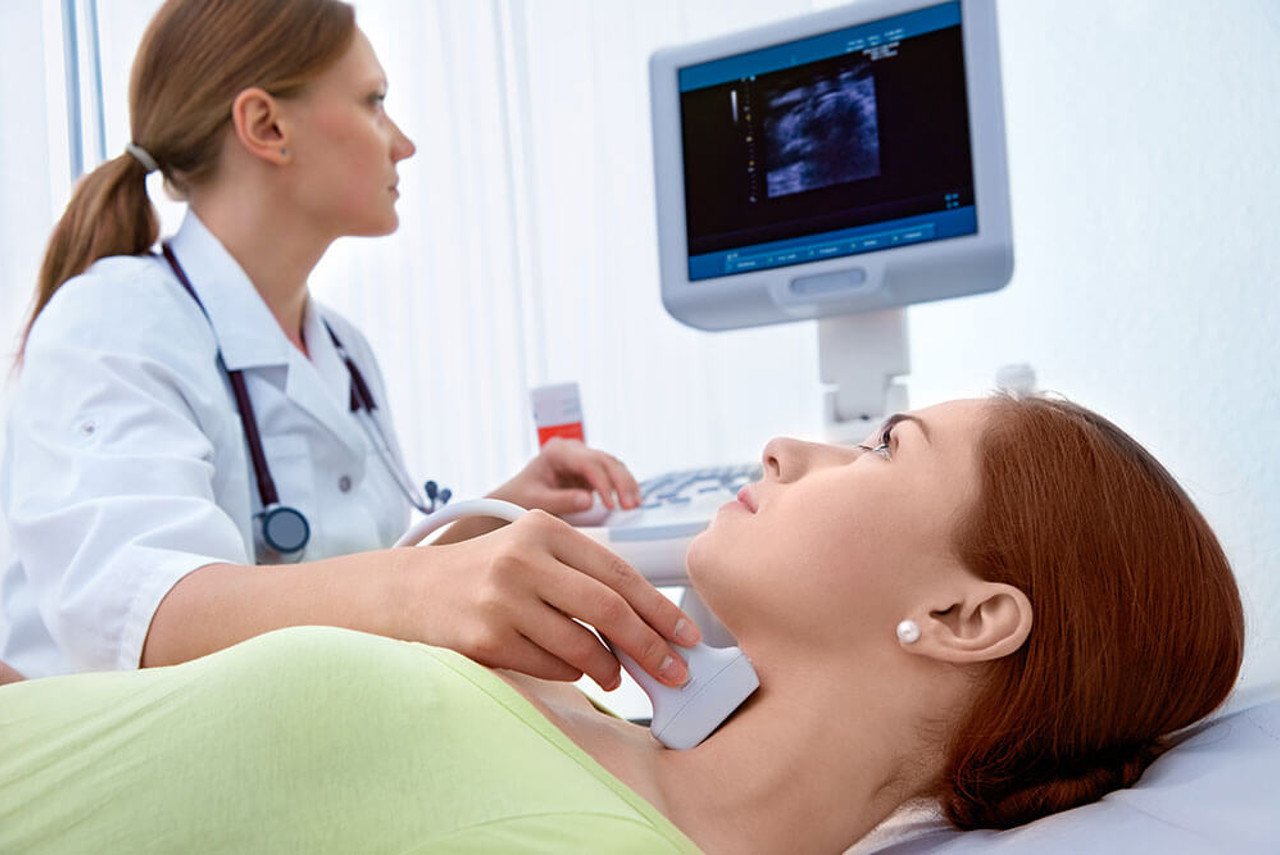
The sonographer will move the ultrasound transducer around the area of interest using a warmed gel and may ask you to move into several different positions during the scan. A small amount of pressure will be applied to the area as the sonographer moves the transducer across the body to capture the images.
-
Vascular ultrasound can take up to 2 hours.
-
Your ultrasound examination will be performed by a sonographer who is a specially trained medical professional.
The images captured by the technicians are viewed by a radiologist who will provide a report to your referring doctor.
How much will it cost?
Fees for radiology procedures will vary depending on a variety of factors. We will advise you about the cost of your service at the time of booking but if you do have any questions, contact us and one of our team will be happy to help with your query. You can read more about our billing information here.
How do I access my images?
At Queensland X-Ray, we provide our patients with their images and results online. To access your images and results, you’ll need to register for an account when you visit one of our practices. If you’ve already registered, you can access the Patient Portal here.
Related procedures
Find another service
Locations providing this service
-
-
189 Lake St
See practice details
Cairns City -
144 Lake Street
See practice details
Cairns City -
318 Mulgrave Road
See practice details
Westcourt
-
-
-
16-24 Weippin Street
See practice details
(Opposite Redland Hospital)
Cleveland -
105 City Road
See practice details
Beenleigh -
Bowen Hills Medical Specialist Centre
See practice details
Suite 3, 16 Thompson Street
Bowen Hills -
14 Grand Plaza Drive
See practice details
Browns Plains -
Ground Floor
See practice details
Rickey Street
Capalaba -
43 Wynyard Street
See practice details
Cleveland -
342 Old Cleveland Road
See practice details
Coorparoo -
Lower Lobby Level
See practice details
Newdegate Street
Greenslopes -
1 Wembley Road
See practice details
Cnr Wembley and Kingston Roads (Service Road)
Logan Central -
589 Logan Road
See practice details
Greenslopes -
62 Bryants Road
See practice details
Shailer Park -
Ground Floor
See practice details
309 Mains Road (Cnr Elva Street)
Sunnybank -
4th Floor
See practice details
32 Raymond Terrace
South Brisbane -
301 Vulture Street
See practice details
South Brisbane -
30 Health Care Drive
See practice details
Springfield Central -
Cnr of Kessels and Troughton Roads
See practice details
Coopers Plains -
Level 1, Sunnybank Private Medical Centre
See practice details
245 McCullough Street,
Sunnybank -
Ground Floor
See practice details
32 Morrow Street
Taringa -
89 Tingal Rd
See practice details
Wynnum -
1437 Logan Road
See practice details
(access via Gowrie Street)
Mount Gravatt
-
-
-
21a/I Domain Central
See practice details
103 Duckworth Street
Garbutt -
Clinical Practice Building
See practice details
James Cook Drive (cnr MT Stuart Street)
Douglas -
Fairfield Homemaker Centre
See practice details
1 D'Arcy Drive
Idalia -
9-13 Bayswater Road
See practice details
Hyde Park -
Mercy Centre
See practice details
25 Fulham Road (access via Diprose Street)
Pimlico -
7/50 North Shore Boulevard
See practice details
Burdell -
Hutchinson Builders Centre Level 2
See practice details
26 Graham Murray Place Railway Estate
Townsville City
-
-
-
Ground Floor
See practice details
14 Hill Street
Southport -
Airport Central
See practice details
1 Eastern Avenue
Bilinga -
GC North Medical Hub
See practice details
502 Hope Island Rd
Helensvale -
Queen Street Village
See practice details
127 Queen Street
Southport -
23 Nexus Way
See practice details
Ground Floor
Southport
-
-
-
73 Highfields Road
See practice details
Highfields -
127 Russell Street
See practice details
Toowoomba City -
280 North Street
See practice details
Toowoomba City -
St Vincent's Hospital
See practice details
Entrance 6, Ground Floor, Herries St
East Toowoomba -
51 Wood Street
See practice details
Warwick -
677 Ruthven Street
See practice details
South Toowoomba
-
-
-
76 Willetts Road
See practice details
North Mackay
-