In 2009, Queensland X-Ray installed the first private PET/CT in the southern hemisphere, and since then has installed additional scanners across the state.
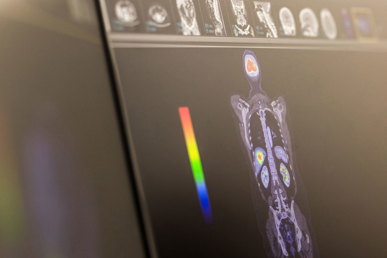
PET stands for Positron Emission Tomography. It uses a small amount of radioactive material, known as a tracer, which is injected into your body and is absorbed into your organs and tissues. CT (Computed Tomography) is a type of X-ray. By combining the two scans, this modality has the ability to detect physiological activity and structural changes of abnormalities or lesions. PET/CT has proven to be extremely sensitive for detecting early stages of cancer and disease, and can localise smaller lesions that may be undetectable by other imaging procedures or CT alone.
PET/CT information can be used to determine what combination of surgery, radiation therapy and chemotherapy is most likely to be successful in managing a patient’s cancer treatment. Additionally, PET/CT can help to monitor the effectiveness of therapy and assist in planning for surgery and radiotherapy.
PET/CT Scan FAQs
-
We use positron emission tomography (PET) imaging, a form of nuclear medicine imaging, combined with CT scanners to provide unparalleled structural detail as well as insight into physiological behaviours within the body, which assists with:
- the more accurate diagnosis of several diseases such as cancer and heart disease
- identifying areas of abnormal activity that can be associated with cancer cells
- assessing changes in blood flow and activity affecting brain and heart tissue
- determining what combination of surgery, radiation therapy and chemotherapy is likely to be the most successful in managing a patient’s cancer
- monitoring the effectiveness of therapy and assisting with a plan for surgery and radiotherapy
By combining functional information from PET imaging with detailed CT anatomy, PET/CT can detect abnormal activity even when there is little or no obvious structural change. This means small tumours may be seen earlier than on other imaging tests or CT alone, supporting a more confident assessment and helping clinicians identify early-stage disease and plan the most appropriate treatment.
-
Correct preparation for a PET/CT is very important to improve the accuracy of the imaging test. Preparation varies between scans and even with the time of your scan. A detailed preparation sheet will be given to you when you make your appointment.
The tracers used in PET have a shelf life of only a few hours, so it’s important you follow the instructions of your doctor and our nuclear medicine technicians closely. If poor preparation forces your appointment to be rescheduled, the tracer ordered for your appointment will expire and this cost may be passed on to you. -
ARRIVAL (15-30MINS)
When you arrive for your appointment, our reception staff will ask that you complete a questionnaire and sign a consent form. In line with your appointment a registered nurse will welcome you through for the procedure. Please be mindful that some patients take longer than others to get settled and your appointment time may vary. We make every effort to keep as closely to appointment times as possible.
TRACER UPTAKE (60-90MINS)
Once you have changed into a gown (if needed) our nurse will get you settled in a private uptake room. They will then check your blood sugar level and place an IV cannula in through which the PET tracer will be injected. You will then be connected to an IV line and be left to relax while the tracer is infused. During this time, you may be permitted to listen to a podcast/audiobook, or watch TV. However, we do not permit any activity that involves holding something to your face (ie watching a movie on your phone or reading a book) as this can affect the quality of the scan. After approximately one hour of rest, we’ll take you through to the PET/CT scanner.
PET/CT IMAGING (20-30MINS)
A CT scanner is doughnut-shaped with a bed passing through the middle. You will be asked to lie on the bed with your arms stretched above your head as the bed moves through the scanner to acquire your images.
The PET part of the scan detects the radiation released from the tracer which has now been taken up throughout your body.
The CT part of the scan images your anatomy and alterations in its structure such as size, shape and other changes related to disease.
The PET and CT images are then combined and used together to diagnose, plan or measure treatment outcomes.
The scan will take between 10 and 25 minutes depending on the reason for your study.POST IMAGING (20-45MINS)
Following your scan you will be offered something to eat and be encouraged to stay well hydrated and use the bathroom as required. We will monitor you for 5-15 minutes following the CT contrast injection and then remove your IV line. During this time we will process your images.
The radioactive tracer remains in your system for a short time following your scan. For this reason, we suggest you limit the time you spend near children and pregnant women for a few hours afterwards. -
Your PET/CT scan will be performed under the supervision of our team of our nuclear medicine specialists, technicians, radiographers and nurses.
The images our technical staff acquire are reviewed by a specialist who will report on the findings. Queensland X-Ray has a wide range of medical imaging specialists. Often PET/CT cases are discussed collectively to offer the most thorough clinical analysis. -
Procedure For staging & restaging of: F-18 FDG Cancer processes GA-68 PSMA Prostate cancer GA-68 DOTATATE Neuroendocrine tumours (NETs) F-18 FET Brain tumours F-18 DOPA Parkinson's disease, NETs, medullary thyroid cancer -
The cost of PET/CT imaging tests can vary depending on a number of factors. We will provide you with the specific price for your PET/CT scan at the time of your booking. If you have any queries about the costs or your eligibility for Medicare, please don’t hesitate to contact us. A member of our team will be more than willing to assist you with your questions.
Watch: Having a PET/CT Scan with Queensland X-Ray
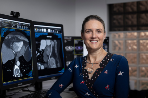
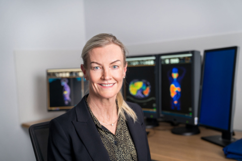
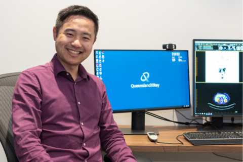
Our PET/CT specialists


How much will it cost?
Fees for radiology procedures will vary depending on a variety of factors. We will advise you about the cost of your service at the time of booking but if you do have any questions, contact us and one of our team will be happy to help with your query. You can read more about our billing information here.
How do I access my images?
At Queensland X-Ray, we provide our patients with their images and results online. To access your images and results, you’ll need to register for an account when you visit one of our practices. If you’ve already registered, you can access the Patient Portal here.