
Queensland X-Ray’s Mount Gravatt practice will soon be home to Queensland’s first Siemens Healthineers NAEOTOM Alpha® photon-counting CT.
Photon-counting CT represents a fundamental advance in CT technology. Unlike conventional energy-integrating detectors, the NAEOTOM Alpha directly counts individual X-ray photons and measures their energy. This enables higher spatial resolution, improved contrast-to-noise performance and spectral information from every scan.
A significant advance in coronary CT angiography
While current high-end conventional CT scanners provide excellent cardiac imaging, photon-counting technology addresses some of their inherent limitations, particularly when imaging small coronary vessels, heavily calcified arteries and coronary stents.
The NAEOTOM Alpha provides in-plane spatial resolution down to 0.11 mm. Combined with 66 ms temporal resolution, this enables exceptionally detailed visualisation of coronary anatomy and small structures, with minimal motion artefact. This is particularly relevant when assessing distal vessels, small-calibre coronary arteries, plaque and complex coronary anatomy where conventional CT may be limited by spatial resolution and artefact.
Addressing the challenge of coronary calcium
Heavy coronary calcification remains one of the limitations of CTCA. Calcium blooming can artificially increase the apparent severity of stenosis and obscure the true vessel lumen. The NAEOTOM Alpha’s Quantum PURE Lumen spectral reconstruction technique is designed to remove the contribution of calcium from coronary CT images and improve visualisation of the underlying lumen.
Improved assessment of coronary stents
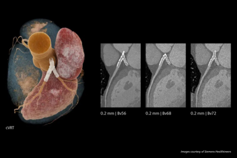
Coronary stents can be challenging to evaluate with conventional CT because of blooming, beam-hardening and metal artefacts, which can obscure the stent lumen and limit assessment for in-stent restenosis. The NAEOTOM Alpha's 0.2 superior spatial resolution provides substantially finer depiction of stent structures and the lumen within the stent, supporting more confident assessment of stent patency and possible in-stent restenosis.
Beyond coronary imaging
The benefits of photon-counting CT extend across a broad range of examinations, with particular value where fine anatomical detail, low-contrast structures or tissue characterisation are important to the diagnosis.
Oncology
Higher-resolution imaging can improve the visualisation of small lesions and subtle anatomical detail, while spectral imaging provides additional information about how lesions enhance following contrast administration. Together, these capabilities may assist with lesion detection, characterisation and assessment of treatment response.
Pulmonary imaging
The increased spatial resolution of photon-counting CT can improve visualisation of small pulmonary nodules and fine lung structures. Lower image noise can also support image quality at lower radiation doses, which is particularly relevant for patients requiring surveillance or repeated chest imaging.
Neurological imaging
Higher spatial resolution can improve visualisation of small cerebral vessels and fine anatomical structures, supporting assessment of conditions such as intracranial vascular disease and other subtle neurological abnormalities.
Musculoskeletal imaging
The ability to acquire very high-resolution images provides detailed assessment of bone, joints and small cortical abnormalities. This can be particularly useful when evaluating subtle fractures, complex anatomy or small osseous lesions. Spectral imaging can also help reduce the impact of artefact from metallic implants, improving assessment of the surrounding anatomy.
Abdominal imaging
Spectral imaging can provide additional information about contrast enhancement and tissue characteristics, which may assist in the detection and characterisation of abdominal lesions and vascular pathology. This additional information is acquired as part of the CT examination, without requiring a separate scan.